1.The Compound Light Microscope
Commonly binocular (two eyepieces), the compound light microscope, combines the power of lenses and light to enlarge the subject being viewed.
Typically, the eyepiece itself allows for 10X or 15X magnification and when combined with the three or four objective lenses, which can be rotated into the field of view, produce higher magnification to a maximum of around 1000X generally.
The compound light microscope is popular among botanists for studying plant cells, to view bacteria and parasites as well as a variety of human/animal cells. It is a useful microscope in forensic labs for identifying drug structures.
Compound light microscopes are one of the most familiar of the different types of microscopes as they are most often found in science and biology classrooms. They are readily available and are inexpensive.
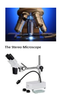
Its also called a dissecting microscope, has two optical paths at slightly different angles allowing the image to be viewed three-dimensionally under the lenses.
Stereo microscopes magnify at low power, typically between 10X and 200X, generally below 100x.
Uses for this type of microscope include looking at surfaces, microsurgery, and watch making, plus building and inspecting circuit boards. Also allow students to observe plant photosynthesis in action.
The Digital Microscope
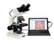
The digital microscope, it uses the power of the computer to view objects not visible to the naked eye.
It connects to a computer monitor via a USB cable, much like connecting a printer or mouse. The computer software allows the monitor to display the magnified specimen. Moving images can be recorded or single images captured in the computer’s memory.
An advantage of digital microscopes is the ability to email images, as well as comfortably watch moving images for long periods.
The popularity of the digital microscope has increased at schools and among hobbyists.
The USB Computer Microscope
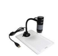
It is among the different types of microscopes, can be used on almost any object and requires no preparation of the specimen. It is essentially a macro lens used to examine images on a computer screen plugged into its USB port.
However, the magnification is restricted and is not comparable to your standard compound light microscope at only up to 200X with a relatively small depth of field.
The Pocket Microscope
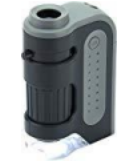
It is used by scientists for hand-held imaging of a variety of specimens/objects in the field or in the laboratory. It is small, durable and portable with a magnification ranging from 25x to 100x.
The Electron Microscope
The Electron Microscope (EM) is a powerful microscope available and used today, allowing researchers to view a specimen at nanometer size. There are two types;
The transmission electron microscope (TEM), the first type of EM, is capable of producing images 1 nanometer in size. The TEM is a popular choice for nanotechnology as well as semiconductor analysis and production.
A second type of electron microscope is the scanning electron microscope(SEM)are approximately 10 times less powerful than TEMs, they produce high-resolution, sharp, black and white 3D images.
They both have practical applications in such fields as biology, chemistry, gemology, metallurgy and industry as well as provide information on the topography, morphology, composition and crystallographic data of samples.
The Scanning Probe Microscope (SPM)
Among the different types of microscopes and microscopy techniques, scanning probe microscopy is used today in academic and industrial settings for those sectors involving physics, biology and chemistry. These instruments are used in research and development as standard analysis tools.
Images are highly magnified and are observed as three-dimensional-shaped-specimens in real time. SPMs employ a delicate probe to scan the surface of the specimen eliminating the limitations that are found in electron and light microscopy.
Titany answered the question on
September 10, 2021 at 11:31